

Photoelectron Spectroscopy
Photoelectron spectroscopy measures the kinetic energies, emission directions, and sometimes spins or coincident products of electrons released when photons ionize a target. The target may be an isolated atom, a molecule, a cluster, a liquid microjet, or a solid surface. What is observed is not a list of pre-existing electron energies. Each spectral channel connects an initial -electron state to a particular state of the residual -electron system plus an outgoing continuum electron.
Energy conservation locates an ionization channel; its matrix element controls the yield and angular pattern. The focus here is conventional one-photon ionization of isolated targets, with the reference-energy and transport distinctions needed for comparison with solids. A measured removal energy is not generally an orbital eigenvalue, and a peak area is not an orbital population.
Required background. Transition rates supplies golden-rule and continuum normalization; molecular orbitals supplies the orbital approximation used for initial assignments. For momentum mapping in solids, see angle-resolved photoemission.
What the Experiment Measures
Section titled “What the Experiment Measures”An ideal experiment has four conceptual stages:
- A source prepares photons with known energy, bandwidth, propagation direction, and polarization.
- The target absorbs a photon and enters one or more ionization channels.
- The outgoing electron propagates through the residual potential and, for condensed targets, through the material and surface.
- An analyzer maps accepted electron trajectories to kinetic energy and a detector records counts.
The minimally differential observable is an energy spectrum,
Angle-resolved detection measures, schematically,
and coincidence experiments may additionally resolve an ionic mass, ionic internal state, fragment momentum, or a second electron.
Counts are not yet a cross section. A useful measurement model is
Here is an integrated photon-flux factor, represents target exposure, is the energy- and angle-dependent acceptance, is the resolution and response kernel from true to measured , and is background. Absolute cross sections require these factors to be calibrated. Relative band areas require at least that their variation across the compared channels be controlled.
A family of methods
Section titled “A family of methods”The photon energy and resolved variables define several overlapping experimental regimes:
| Method | Typical emphasis | Information carried |
|---|---|---|
| UV photoelectron spectroscopy (UPS) | Valence ionization | Molecular valence channels, work functions, valence bands |
| X-ray photoelectron spectroscopy (XPS or ESCA) | Core ionization | Element and chemical-state sensitivity, surface composition |
| Angle-resolved photoemission (ARPES) | Energy and emission angle from ordered solids | Occupied band dispersion and removal spectral weight |
| Photoelectron imaging | Energy and angle, often for gas-phase species | Channel-resolved angular distributions and dynamics |
| Threshold photoelectron spectroscopy | Near-zero-kinetic-energy electrons | Accurate ionization thresholds and ionic-state structure |
| Photoelectron–photoion coincidence | Electron plus mass-selected ion | State-selected ions, fragmentation, and internal-energy flow |
These names describe experimental emphasis, not different conservation laws. UPS and XPS both measure photoemitted electrons. A sharp boundary based only on photon energy is conventional rather than fundamental.
Every line labels a final channel
Section titled “Every line labels a final channel”Let the prepared target state be and let denote a final channel with residual ionic state and an outgoing electron of asymptotic momentum . The channel-resolved transition amplitude is
where specifies photon polarization and is the light–matter interaction in a declared gauge and approximation.
The amplitude contains the many-electron overlap, dipole selection, continuum distortion, partial-wave interference, and channel coupling. Two ionic states at similar binding energy can have very different intensities. Conversely, one nominal one-particle removal can distribute intensity among a main line and several satellites.
Ionization Energies of Isolated Targets
Section titled “Ionization Energies of Isolated Targets”For an isolated target initially at rest, nonrelativistic energy conservation for channel is
Define the state-to-state ionization energy
Then
Recoil is often negligible at routine molecular UPS resolution but is a real part of precision atomic and molecular measurements. The electron and ion share momentum, so the ion carries
Photon momentum, target thermal motion, and fragment recoil may also matter in high-resolution or coincidence work.
Binding energy versus ionization energy
Section titled “Binding energy versus ionization energy”For an isolated atom or molecule, a reported band binding energy is often used operationally for
after recoil and calibration conventions have been handled. It then equals a state-to-state ionization energy for the channel being measured. The phrase ionization energy is safer when discussing the threshold between well-defined neutral and ionic states.
The first adiabatic ionization energy is the lowest energy required to reach the ground rovibronic state of the ion from the ground rovibronic state of the neutral. Selection rules can suppress access to this threshold in a particular experiment; they do not redefine the state-to-state ionization energy. Excited ionic states produce higher ionization bands.
Vertical and adiabatic molecular ionization
Section titled “Vertical and adiabatic molecular ionization”Let denote nuclear geometry. For electronic ionic state , a fixed-geometry or vertical electronic ionization energy from neutral state is
The vertical value most often quoted uses the equilibrium geometry of the neutral:
An adiabatic value compares separately relaxed minima and includes the declared zero-point contributions:
The adiabatic threshold and the maximum of a broad photoelectron band need not coincide. A band maximum depends on Franck–Condon factors, initial temperature, rotational structure, channel-dependent matrix elements, lifetimes, and instrumental response.
Vibronic channels
Section titled “Vibronic channels”Within a Born–Oppenheimer description, ionization can populate many ionic vibrational states:
Under a Condon-like approximation, a band intensity contains a nuclear overlap,
multiplied by an electronic photoionization factor that can itself vary with energy and channel. A resolved progression measures ionic vibrational spacings and geometry change. It is not merely instrumental broadening.
The nuclear geometry is effectively frozen during the sudden electronic transition, not after it. Broadband coherent preparation can create an ionic wavepacket that subsequently vibrates. Narrowband excitation resolved to a single bound ionic vibrational level instead prepares a stationary level; tracing over an entangled photoelectron can also remove ionic coherence. Populated unstable ionic states may predissociate, isomerize, or fragment. Coincidence detection is often needed to associate an electron band with one of those outcomes.
Photodetachment
Section titled “Photodetachment”Removing an electron from an anion is usually called photodetachment. The same channel energy balance applies, but the residual species may be neutral:
The lowest threshold is the electron binding energy of the anion, equal to the electron affinity of the corresponding neutral when both refer to the appropriate ground states. Near threshold, the absence of an attractive residual Coulomb tail makes Wigner-law behavior especially transparent.
Binding-Energy Reference Zeros
Section titled “Binding-Energy Reference Zeros”The formula is incomplete until both energies have declared zeros. This matters acutely when an electron travels from a solid sample into an analyzer with a different work function.
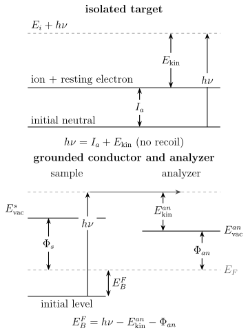
For an isolated target, a channel threshold and its electron kinetic energy add to when recoil is neglected. For a conducting sample in electrical contact with an analyzer, Fermi levels align and the calibrated analyzer work function enters the usual Fermi-referenced binding-energy equation. Mixing these reference conventions creates work-function-sized errors.
Grounded conducting samples
Section titled “Grounded conducting samples”Suppose a conducting sample and analyzer are in electrical equilibrium, so their Fermi levels align. If the analyzer reports kinetic energy relative to its own vacuum level, the conventional Fermi-referenced binding energy is
The analyzer work function is part of the calibrated instrument energy scale. The sample work function does not appear as an additional subtraction in this equation: the contact potential between sample and analyzer accounts for the difference between their vacuum levels.
Relative to the sample vacuum level,
This vacuum-referenced quantity is useful for connecting a valence onset to an ionization energy, but it requires the sample work function and a clear surface condition.
Work function and secondary-electron cutoff
Section titled “Work function and secondary-electron cutoff”The sample work function is
In UPS, one can infer it from the full kinetic-energy width between the Fermi edge and secondary-electron cutoff, with the applied sample bias and analyzer convention included. A commonly used ideal relation is
The bias shifts measured kinetic energies so that low-energy electrons clear the analyzer threshold. It must be included consistently; subtracting it twice is a common failure.
Insulators, charging, and referencing
Section titled “Insulators, charging, and referencing”An insulating specimen may accumulate positive charge as electrons leave. A uniform electrostatic shift approximately moves all photoelectron features by
Real specimens can charge differentially, producing position-dependent shifts and broadening. A single post hoc rigid offset cannot repair a nonuniform potential distribution.
Binding-energy referencing and instrument calibration are separate tasks:
- calibration establishes the analyzer energy scale using traceable reference lines and a documented procedure;
- referencing identifies the physically relevant zero for the specimen;
- charge correction estimates specimen-induced electrostatic shifts.
Assigning an arbitrary contaminant peak a universal binding energy can hide charging, chemical shifts, or an incorrect model. Report the reference material, line, assumed value, electrical contact, neutralization method, and applied correction.
Chemical shifts are total-energy shifts
Section titled “Chemical shifts are total-energy shifts”A core-level chemical shift compares channel energies in different chemical environments. It can reflect:
- initial-state charge redistribution and electrostatic potential;
- changes in screening and orbital relaxation after core-hole creation;
- multiplet and spin–orbit structure;
- local bonding, polarization, and final-state correlation;
- referencing, charging, and surface band bending.
It is therefore not a direct meter of one atomic partial charge. Interpreting a shift requires comparison within a controlled chemical series and an explicit electronic-structure or screening model.
From Ideal Channels to a Spectrum
Section titled “From Ideal Channels to a Spectrum”For weak monochromatic light, a channel-resolved differential photoionization cross section has the golden-rule structure
The exact prefactor depends on unit system, photon-state normalization, continuum normalization, and the chosen form of the interaction operator. Physical cross sections are gauge independent in an exact calculation; disagreement between length and velocity forms in a truncated calculation can expose incomplete wavefunctions or inconsistent operators. A measured spectrum then convolves this cross section with source bandwidth, target distributions, analyzer response, and electron transport.
Resolution and line shape
Section titled “Resolution and line shape”If statistically independent broadening contributions are approximately Gaussian, their standard deviations add in quadrature:
The same is not true for arbitrary full widths or non-Gaussian profiles. Lorentzian lifetime widths add linearly under convolution, and a Gaussian–Lorentzian convolution gives a Voigt profile. Vibrational envelopes, unresolved multiplets, asymmetric metallic line shapes, and inelastic-loss tails require physical models rather than a larger generic width.
Line Shapes and Broadening develops these distinctions.
Analyzer acceptance and transmission
Section titled “Analyzer acceptance and transmission”An analyzer accepts a finite energy and angular range. Its transmission can vary strongly with pass energy, lens mode, aperture, and kinetic energy. Thus an integrated band area is more accurately written
Comparing raw areas from different kinetic energies, analyzer settings, or photon energies is not a population measurement. Photoionization cross sections and asymmetry parameters also vary with photon energy.
Electron transport and surface sensitivity
Section titled “Electron transport and surface sensitivity”In a condensed target, an electron created at depth and emitted at angle from the surface normal is attenuated approximately as
where is an inelastic mean free path in a simple planar model. Elastic scattering, surface roughness, anisotropy, diffraction, and finite acceptance modify this expression.
The measured signal is therefore depth weighted. Changing kinetic energy or takeoff angle changes the sampling depth. “Surface sensitive” does not mean “only the top atomic layer,” and a quoted information depth should state its criterion and transport model.
Backgrounds and energy losses
Section titled “Backgrounds and energy losses”Photoelectrons that undergo inelastic scattering can populate a structured background below their original kinetic energy, equivalently at higher apparent binding energy. Plasmon losses, interband excitations, and secondary electrons are physical signals, not merely detector noise.
Background subtraction is a model choice. Shirley, Tougaard, polynomial, and locally fitted backgrounds encode different assumptions. Peak areas and satellite intensities should be reported with sensitivity to plausible background models.
Koopmans-Style Interpretation
Section titled “Koopmans-Style Interpretation”Canonical Hartree–Fock orbitals provide a useful first assignment language. If an electron is removed from occupied spin-orbital and every remaining orbital is frozen, Koopmans’ identity gives
Within that constrained Hartree–Fock model, this is an exact determinant energy identity. It is not an exact statement that a measured peak equals an orbital eigenvalue.
Why it can organize a valence spectrum
Section titled “Why it can organize a valence spectrum”For a weakly correlated closed-shell molecule:
- low-lying ionic states may be dominated by one-hole configurations;
- occupied canonical orbitals can supply symmetry and bonding labels;
- the ordering of may track main-band ordering;
- Franck–Condon progressions can reveal how each ionic surface differs from the neutral surface.
This makes “ionization from orbital ” useful shorthand when its approximation is understood. A careful assignment says that an ionic state has dominant one-hole character associated with orbital , not that the instrument measured the orbital eigenvalue directly.
Relaxation, correlation, and ionic-state mixing
Section titled “Relaxation, correlation, and ionic-state mixing”After removal, the remaining electrons relax in the altered potential. When optimization stays within an admissible ionic variational space containing the frozen determinant,
so orbital relaxation lowers the ionized determinant energy relative to the frozen construction. Correlation changes both neutral and ionic energies and can mix several one-hole and multielectron configurations.
One can organize the corrections schematically:
The signs and partition of these corrections depend on definitions and method. The individual correction terms are not separately measurable. For inner-shell holes, relaxation can be large. For open shells, removal from one determinant orbital can project onto several ionic multiplets.
Kohn–Sham eigenvalues
Section titled “Kohn–Sham eigenvalues”Kohn–Sham orbitals belong to an auxiliary system constructed to reproduce a density. Under the usual exact ground-state assumptions, the highest occupied exact Kohn–Sham eigenvalue has a special relation to the first ionization energy. This does not make every occupied eigenvalue of an approximate functional an exact photoelectron binding energy.
Generalized Kohn–Sham eigenvalues can be useful empirical assignment tools, but comparisons must state the functional, orbital convention, geometry, relativistic treatment, and whether energies were obtained from orbital eigenvalues, total-energy differences, quasiparticle theory, or an explicit ionized-state calculation.
A practical hierarchy
Section titled “A practical hierarchy”For peak positions, increasingly complete models include:
- frozen-orbital Hartree–Fock estimates;
- separately optimized neutral and ionic total-energy differences;
- correlated ionization-potential or electron-propagator methods;
- vibronic, relativistic, and continuum-coupled calculations;
- explicit simulation of instrumental and transport response.
Higher on this list does not automatically mean better. A calculation must converge the relevant states, represent the continuum, and match the measured observable. A highly correlated bound-state energy with a crude continuum can predict peak positions well while failing angular distributions.
Molecular-Orbital Information
Section titled “Molecular-Orbital Information”A photoelectron spectrum contains genuine orbital information, but only through state-to-state removal amplitudes and a forward model.
Dyson orbitals
Section titled “Dyson orbitals”For normalized initial state and ionic state , define the Dyson spin-orbital
It is the overlap left after electron coordinates have been integrated out. Different ionic states have different Dyson orbitals, even when a one-orbital picture assigns them to the same nominal shell.
In a single-channel dipole approximation that neglects additional continuum correlation and retains the direct removal contribution, the amplitude can often be organized as
where is a continuum electron state distorted by the ionic channel. This compact expression is powerful, but it is an approximation to a fully antisymmetrized many-electron scattering amplitude. Interchannel coupling and correlation in the continuum may not factorize.
Pole strength and one-electron character
Section titled “Pole strength and one-electron character”The Dyson-orbital norm
is often called a pole strength or spectroscopic factor in an appropriate Green-function convention. A value near one indicates strong one-electron removal character for that channel. Correlation can distribute strength across many ionic states so that individual values are smaller. For one normalized, individually resolved ionic state, . Summing the norms over a complete set of residual states gives , not one; unresolved spin or other degenerate channels must be counted explicitly.
Measured integrated intensity is not generally equal to :
It also contains continuum matrix elements, photon-energy dependence, polarization, molecular orientation, degeneracy, analyzer acceptance, and transport. Pole strength is a property of the many-body overlap; observed area is a property of the full experiment.
Main lines and satellites
Section titled “Main lines and satellites”In a one-hole picture, a main line connects strongly to an ionic state dominated by deleting one occupied orbital. Correlation can create additional channels:
- shake-up: ionization accompanied by a bound excitation of the residual system;
- shake-off: ionization accompanied by emission of another electron into the continuum;
- multiplet splitting: several ionic terms formed by coupling the hole to open-shell angular momenta;
- screened and unscreened final states: distinct many-body responses to a hole, especially in solids and transition-metal compounds;
- plasmon or other loss structures: extrinsic, intrinsic, or interfering energy-loss processes.
A weak satellite need not be contamination, and a strong satellite need not represent a highly occupied orbital. Satellite positions and intensities are among the most direct signs that the one-hole approximation is incomplete.
Cross-section zeros and missing bands
Section titled “Cross-section zeros and missing bands”Even a channel with substantial Dyson norm can become weak if its dipole matrix element is small. A radial dipole integral can pass through zero as photon energy varies, producing a Cooper minimum in a partial photoionization cross section. Symmetry, orientation averaging, destructive partial-wave interference, and detector geometry can likewise suppress a band.
Absence from one spectrum is therefore not proof that an orbital or ionic state does not exist. Varying photon energy and polarization is a central assignment test.
Extended systems and spectral functions
Section titled “Extended systems and spectral functions”For a translationally ordered solid, angle-resolved photoemission can be related under a sudden, single-particle-like treatment to
convolved with resolution and supplemented by background and surface effects. Here is the full one-electron spectral function, including removal and addition, and is the equilibrium Fermi factor, with measured relative to the chemical potential in the chosen energy units. The occupied removal spectrum is ; if is used instead, it must not be multiplied by again.
This formula is a useful map, not a license to identify intensity directly with . Matrix-element zeros, finite escape depth, surface states, uncertainty, final-state structure, domains, and detector acceptance all matter. Spectral Functions derives the Lehmann representation, quasiparticle poles, and incoherent weight.
Photoelectron Angular Distributions
Section titled “Photoelectron Angular Distributions”Energy conservation says which channel is open. The angular distribution reveals how photon polarization, target symmetry, continuum angular momenta, and scattering phases combine within that channel.
Random targets and linearly polarized light
Section titled “Random targets and linearly polarized light”For one-photon ionization of a randomly oriented target in the electric-dipole approximation, with no resolved electron spin, the laboratory-frame distribution has the form
where
The angle is measured from the linear-polarization axis. Positivity imposes
Three limiting patterns are useful:
| Angular character | Relative pattern | |
|---|---|---|
| Strongly parallel | ||
| Isotropic in this laboratory frame | constant | |
| Strongly perpendicular |
Because
the distribution integrates to . An analyzer at one angle samples a -dependent fraction of the total cross section.
At the magic angle
the dipole anisotropy term vanishes. This can simplify comparisons, but only when the target, polarization, acceptance, and dipole assumptions fit the formula.
Partial waves and dipole selection
Section titled “Partial waves and dipole selection”In a central-field one-electron description, for initial orbital angular momentum , an electric-dipole transition produces continuum channels
Their radial amplitudes and scattering phases interfere. The total cross section depends on squared amplitudes, while also depends on relative phases. Thus two models can predict similar total yield but different angular patterns.
A Cooper minimum occurs when a relevant radial dipole matrix element changes sign through zero. Near the minimum:
- the partial cross section can become small;
- a normally subdominant channel can control the angular pattern;
- can vary rapidly;
- correlation and relativistic channel coupling become conspicuous.
This is scattering-phase information encoded in photoemission.
Molecules and molecular-frame distributions
Section titled “Molecules and molecular-frame distributions”For a randomly oriented molecule, orientation averaging reduces a one-photon dipole distribution to the same form. That single number does not retain the complete molecular-frame pattern.
If the molecular axis is known through alignment, orientation, or coincident fragment recoil, the differential cross section can depend on several angles and polarization tensors. Molecular-frame photoelectron angular distributions can expose nodal structure, shape resonances, multicenter interference, and the symmetry of the residual ion.
Reconstructing an “orbital image” from such data requires:
- a state-specific ionic channel;
- a continuum scattering model;
- an orientation and recoil model;
- a gauge and phase convention;
- treatment of finite resolution and detector acceptance.
The reconstructed object is a model-dependent transition amplitude, often closely related to a Dyson orbital, not a photograph of a stationary electron cloud.
Beyond the simple anisotropy formula
Section titled “Beyond the simple anisotropy formula”Additional angular coefficients are required for:
- oriented or aligned targets;
- circular or elliptical polarization;
- spin-resolved detection;
- nondipole electric-quadrupole and magnetic-dipole contributions;
- multiphoton ionization;
- chiral photoelectron circular dichroism;
- coincident electrons or fragments.
At higher photon energies, terms odd in the photon propagation direction can produce forward–backward asymmetry. Fitting such data with only can fold nondipole physics into a misleading dipole parameter.
Thresholds, Resonances, and Scattering
Section titled “Thresholds, Resonances, and Scattering”The emitted electron is a scattering particle in the field of the residual target. Photoionization is therefore a bound-to-continuum scattering problem, not merely a bound-state energy difference.
Wigner threshold law
Section titled “Wigner threshold law”For a short-range residual interaction and an outgoing partial wave , the near-threshold partial cross section behaves as
or, since ,
The lowest allowed partial wave usually dominates sufficiently close to threshold. For photodetachment into an wave, ; for a wave, .
The assumptions matter. A residual positive ion has an attractive Coulomb tail, for which the simple short-range exponent is modified. Polarization potentials, dipole-bound states, virtual states, resonances, and channel coupling can also alter the observed onset.
Direct ionization and autoionization
Section titled “Direct ionization and autoionization”In direct photoionization, the photon couples directly to an open ion-plus-electron channel. A photon can also excite a discrete state embedded in a continuum, which then decays by ejecting an electron. Interference between direct and resonant paths can produce a Fano profile:
Here and locate and broaden the resonance, while encodes the relative resonant and direct amplitudes in the ideal one-continuum model. The asymmetric profile is amplitude interference, not two unresolved positive peaks.
Autoionization and Auger decay are related but distinct labels. In autoionization, an excited state above an ionization threshold ejects an electron through electron correlation. In an Auger process, a vacancy is filled while another electron is emitted. The latter electron kinetic energy is primarily set by electronic level differences and is not shifted one for one with the exciting photon energy.
Continuum boundary conditions
Section titled “Continuum boundary conditions”The state in a photoionization matrix element carries incoming-wave scattering boundary conditions in a common bra convention; the photoemission amplitude uses its bra to project onto an outgoing asymptotic channel. For a real, time-reversal-invariant spinless Hamiltonian, : complex conjugation also reverses momentum. Replacing by without this transformation is not a harmless change of notation.
The partial-wave form is schematically
The scattering phase shifts control angular interference and resonance behavior. A plane-wave final state discards these phases and can fail badly at low energy, near shape resonances, around Cooper minima, or in multicenter molecular potentials.
Time-reversed photorecombination
Section titled “Time-reversed photorecombination”Microscopic reversibility relates state-resolved photoionization to the time-reversed radiative recombination process,
Detailed-balance factors account for phase-space and degeneracy differences. The same dipole amplitudes and continuum phases appear in both directions. High-harmonic recombination also probes a related time-reversed matrix element, although strong-field preparation and propagation add substantial physics.
Scattering inside the target
Section titled “Scattering inside the target”For molecules and solids, the outgoing wave can scatter from neighboring centers before detection. This produces:
- molecular shape resonances;
- multicenter interference;
- photoelectron diffraction;
- energy-dependent final-state phases;
- elastic deflection and inelastic losses.
The relation to scattering is therefore twofold: scattering theory defines the final continuum state, and actual post-emission transport reshapes the detected distribution.
Worked Examples
Section titled “Worked Examples”Gas-phase channel assignment
Section titled “Gas-phase channel assignment”A He I source supplies
A molecular band has calibrated electron kinetic energy
Neglecting recoil at the quoted precision gives
This number labels the populated ionic channel. Calling it a HOMO energy requires the additional claim that the channel is dominated by removal from the neutral HOMO and that a Koopmans-like interpretation is adequate.
Analyzer-referenced XPS
Section titled “Analyzer-referenced XPS”For monochromated Al radiation, take
If the analyzer reports and its calibrated work function is , then
Subtracting the sample work function again would double count the contact potential convention.
Angular acceptance
Section titled “Angular acceptance”For , the ideal differential intensity along the polarization axis is proportional to
At ,
An infinitesimal detector parallel to the polarization therefore sees ten times the differential intensity of one at :
A real analyzer averages over finite angular acceptance, so its measured ratio is less extreme unless the acceptance integration is included.
Exercises
Section titled “Exercises”Exercise 1: Recoil precision
Section titled “Exercise 1: Recoil precision”A photon of energy ionizes a stationary molecule into a channel with threshold . The measured electron kinetic energy is . Assuming no fragmentation, what recoil energy is implied? Would it be consistent to quote the threshold to while neglecting this term?
Solution
Energy conservation gives
Therefore
The recoil correction is ten times the proposed precision. It cannot be neglected at that level. The rounded numbers in the question do not themselves support precision; photon energy, thermal motion, molecular mass, and calibration must also be consistent with the inferred recoil.
Exercise 2: Vertical versus adiabatic ionization
Section titled “Exercise 2: Vertical versus adiabatic ionization”At the neutral equilibrium geometry, a calculation gives a vertical ionization energy of . Relaxing the ion lowers its electronic energy by relative to that vertical ionic geometry. The ionic zero-point energy exceeds the neutral zero-point energy by . Estimate the adiabatic ionization energy. Why need neither value coincide exactly with the band maximum?
Solution
The relaxed electronic minimum lowers the ionization difference by , while the zero-point difference raises it by :
The adiabatic energy refers to the two lowest rovibronic levels under the stated model. The vertical energy is a fixed-geometry difference. A measured band maximum is selected by the Franck–Condon envelope, rotational and thermal populations, matrix-element variation, linewidths, and instrument response, so it is not defined by either energy alone.
Exercise 3: Work-function conventions
Section titled “Exercise 3: Work-function conventions”A grounded conducting sample is measured with photons of . The analyzer work function is and the measured kinetic energy is . The independently measured sample work function is .
- Find the Fermi-referenced binding energy.
- Find the sample-vacuum-referenced binding energy.
- Explain why the sample work function is absent from the first calculation.
Solution
The Fermi-referenced value is
The vacuum-referenced value is
Electrical contact aligns sample and analyzer Fermi levels. Their unequal work functions create a contact potential that shifts the electron kinetic energy en route to the analyzer. Using the analyzer-referenced kinetic energy and calibrated analyzer work function already includes that shift.
Exercise 4: Koopmans and relaxation
Section titled “Exercise 4: Koopmans and relaxation”An occupied Hartree–Fock orbital has . Reoptimizing the ionic Hartree–Fock determinant lowers its energy by relative to the frozen ion. Correlation and relativistic corrections together raise the predicted ionization energy by relative to the relaxed Hartree–Fock value. Estimate the three successive ionization energies and identify which one Koopmans’ theorem supplies.
Solution
Koopmans’ frozen-orbital value is
Relaxation lowers the ionic total energy and hence the ionization energy:
The stated remaining corrections raise it:
Only the first value is the frozen-orbital Koopmans result. The numerical partition is model dependent; in a real calculation, correlation corrections must be computed for both neutral and ionic states rather than added by rule of thumb.
Exercise 5: Pole strength is not peak area
Section titled “Exercise 5: Pole strength is not peak area”Two ionic channels have Dyson norms and . At one photon energy, the continuum and detector factors multiplying these norms are in the ratio . Under the simplified model , what area ratio is expected? What lesson survives when the factorization itself fails?
Solution
The simplified areas satisfy
The channel with the larger Dyson norm has the smaller observed area because its continuum and detection factor is weaker. More generally, the exact amplitude may not factor into a pole strength times a scalar continuum factor. The robust lesson is that a peak area is not a direct measurement of Dyson norm, occupancy, or electron count.
Exercise 6: Finite angular acceptance
Section titled “Exercise 6: Finite angular acceptance”An analyzer uniformly accepts a cone of half-angle about an axis at angle to the light polarization. Average the dipole angular factor over that cone. Compare parallel and perpendicular cone axes for and .
Solution
Azimuthal averaging about the cone axis gives . With , the remaining average is
Thus the averaged factor is . At , and the ratio is , below the infinitesimal-detector ratio of ten. A symmetric cone centered on the magic angle still cancels the rank-2 term; asymmetric or nonuniform acceptance need not do so.
Exercise 7: Threshold exponents
Section titled “Exercise 7: Threshold exponents”For a short-range residual potential, compare the increase in cross section when the excess energy rises from to for an outgoing wave and for an outgoing wave.
Solution
Wigner’s law gives
For an wave, :
For a wave, :
The lower allowed partial wave rises more slowly but dominates sufficiently close to threshold because higher partial waves are more strongly suppressed. The result must not be applied unchanged to an attractive Coulomb residual potential.
Exercise 8: Total removal strength
Section titled “Exercise 8: Total removal strength”Let annihilate an electron in an orthonormal spin-orbital and define . Show that a complete sum of equals . Why does a small total norm in a selected energy window not imply a violation of particle conservation?
Solution
Completeness in the -electron space gives
The complete sum includes all residual excitations and continuum states, with integrals where required. A finite spectral window can omit substantial removal weight. Measured photoelectron area has additional transition and detector factors, so even a complete area integral is not this sum rule without a justified conversion.
For timed ionization of electronic and nuclear motion, see ultrafast spectroscopy. Partial waves and Coulomb continuum states develop the final-state scattering used here.
References
Section titled “References”- J. Cooper and R. N. Zare, “Angular Distribution of Photoelectrons,” Journal of Chemical Physics 48, 942–943 (1968), doi:10.1063/1.1668742 — dipole anisotropy formula and partial-wave interpretation.
- C. S. Fadley, “X-ray Photoelectron Spectroscopy: Progress and Perspectives,” Journal of Electron Spectroscopy and Related Phenomena 178–179, 2–32 (2010), doi:10.1016/j.elspec.2010.01.006 — core-level photoemission, diffraction, surfaces, and modern extensions.
- U. Fano, “Effects of Configuration Interaction on Intensities and Phase Shifts,” Physical Review 124, 1866–1878 (1961), doi:10.1103/PhysRev.124.1866 — interference between discrete and continuum pathways.
- S. Hüfner, Photoelectron Spectroscopy: Principles and Applications, 3rd ed., Springer, 2003, doi:10.1007/978-3-662-09280-4 — photoemission from atoms, molecules, solids, and surfaces.
- IUPAC, “ionization energy,” Compendium of Chemical Terminology, 5th ed., 2025 — adiabatic and vertical ionization-energy terminology.
- IUPAC, “photoelectron spectroscopy,” Compendium of Chemical Terminology, 5th ed., 2025 — measurement definition and PES, UPS, XPS, and ESCA terminology.
- T. Koopmans, “Über die Zuordnung von Wellenfunktionen und Eigenwerten zu den einzelnen Elektronen eines Atoms,” Physica 1, 104–113 (1934), doi:10.1016/S0031-8914(34)90011-2 — original frozen-orbital relation.
- A. Müller, S. Schippers, A. Aguilar, I. Alvarez, M. E. Bannister, J. Bozek, C. Cisneros, A. M. Covington, G. H. Dunn, M. F. Gharaibeh, G. Hinojosa, S. Ricz, A. S. Schlachter, and R. A. Phaneuf, “Time-Reversal Studies in Photorecombination and Photoionization Experiments with Ion Beams,” AIP Conference Proceedings 680, 191 (2003), doi:10.1063/1.1619696 — state-resolved detailed balance between photoionization and photorecombination.
- NIST Standard Reference Database 20, X-ray Photoelectron Spectroscopy Database, version 4.1 — evaluated binding energies, chemical shifts, Auger energies, and measurement metadata.
- C. M. Oana and A. I. Krylov, “Dyson Orbitals for Ionization from the Ground and Electronically Excited States within Equation-of-Motion Coupled-Cluster Formalism: Theory, Implementation, and Examples,” Journal of Chemical Physics 127, 234106 (2007), doi:10.1063/1.2805393.
- C. J. Powell, “Calibrations and Checks of the Binding-Energy Scales of X-ray Photoelectron Spectrometers,” Journal of Electron Spectroscopy and Related Phenomena 257, 146808 (2022), doi:10.1016/j.elspec.2018.11.007 — analyzer calibration, reference energies, drift, and uncertainty.
- J. W. Rabalais, Principles of Ultraviolet Photoelectron Spectroscopy, Wiley, 1977 — molecular valence ionization, vibronic structure, and angular distributions.
- M. P. Seah, I. S. Gilmore, and S. J. Spencer, “Measurement of Data for and the Development of an ISO Standard for the Energy Calibration of X-ray Photoelectron Spectrometers,” Surface and Interface Analysis 31, 778–795 (2001), doi:10.1002/sia.1115 — multi-line binding-energy calibration procedure.
- C. M. Truesdale, S. H. Southworth, P. H. Kobrin, U. Becker, D. W. Lindle, H. G. Kerkhoff, and D. A. Shirley, “Shape Resonance Phenomena in CO Following K-Shell Photoexcitation,” Physical Review Letters 50, 1265 (1983), doi:10.1103/PhysRevLett.50.1265 — continuum scattering and molecular shape-resonance anisotropy.
- D. W. Turner, C. Baker, A. D. Baker, and C. R. Brundle, Molecular Photoelectron Spectroscopy, Wiley-Interscience, 1970 — foundational gas-phase molecular photoelectron spectroscopy.
- E. P. Wigner, “On the Behavior of Cross Sections Near Thresholds,” Physical Review 73, 1002–1009 (1948), doi:10.1103/PhysRev.73.1002 — threshold laws and the role of long-range product interactions.
- J. J. Yeh and I. Lindau, “Atomic Subshell Photoionization Cross Sections and Asymmetry Parameters: ,” Atomic Data and Nuclear Data Tables 32, 1–155 (1985), doi:10.1016/0092-640X(85)90016-6 — tabulated photon-energy-dependent cross sections and anisotropies.